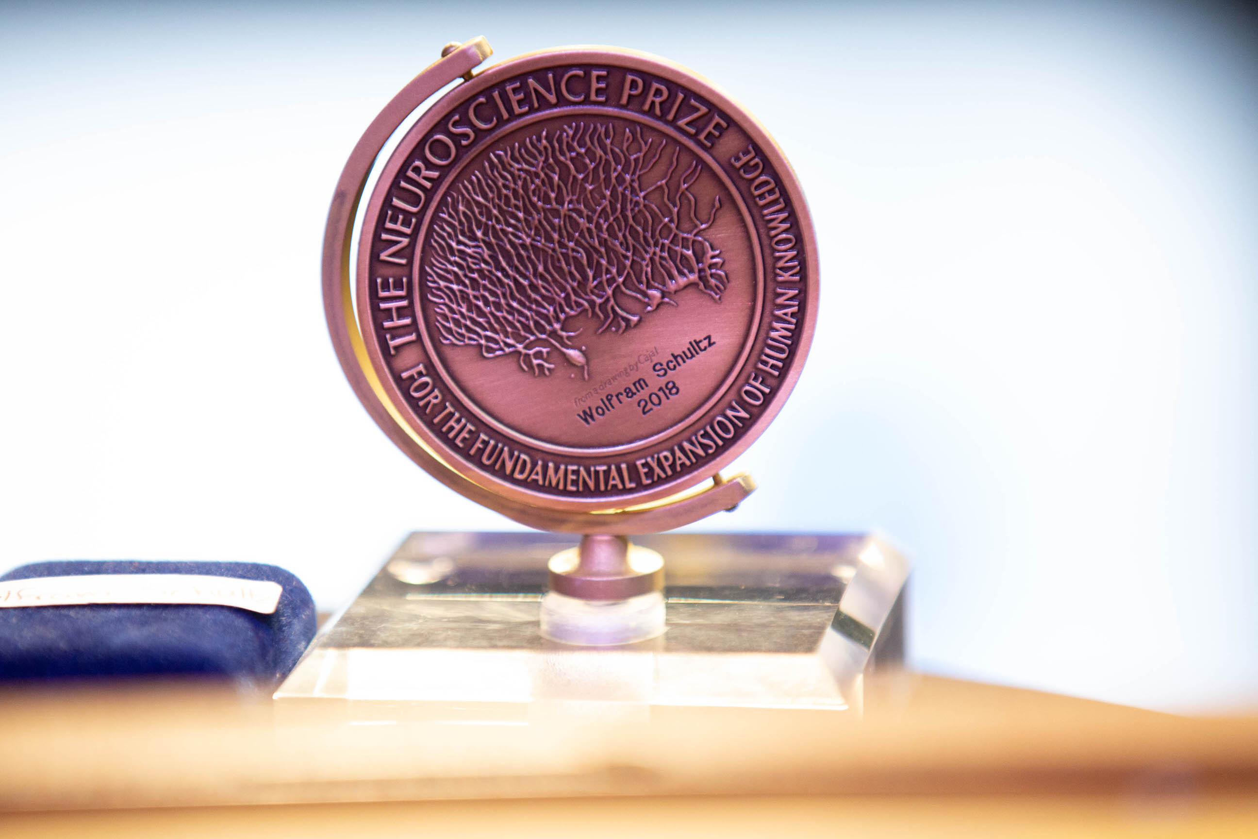
2018 Gruber Neuroscience Prize
Ann M. Graybiel, of the Massachusetts Institute of Technology, Okihide Hikosaka, of the National Institutes for Health’s National Eye Institute, and Wolfram Schultz, of the University of Cambridge, have been pioneers in the study of the structure, organization and functions of the basal ganglia, a group of nuclei (clusters of neurons) located deep within the forebrain. Their groundbreaking work has fundamentally transformed our understanding of the basal ganglia and has led to influential new ideas about how the brain learns and retains new habits and skills, manages movements and processes rewards for learning and decision-making. It has also deepened our understanding of a wide range of neurodegenerative and neuropsychiatric disorders in which the basal ganglia and behavioral control are compromised.
2018 Neuroscience Prize Recipients


Laureate Profile
Ann M. Graybiel, PhD, of the McGovern Institute for Brain Research at the Massachusetts Institute of Technology (MIT), pioneered our understanding of the important role that basal ganglia, a group of nuclei (clusters of neurons) deep within the forebrain, play in a wide range of neurological disorders. At the time she started her research, in the 1970s, most scientists were ignoring that area of the brain. Graybiel, however, persevered. In a groundbreaking experiment, she found that the striatum, the largest nucleus within the basal ganglia, was not a homogenous mass of cells (as was commonly believed at the time). Instead, it had a distinct and sophisticated architecture with column-like modules — dubbed striosomes by Graybiel — that distributed nearly every known neurotransmitter. She also found that striosomes were surrounded by a matrix, which was itself modular. In an extraordinary series of subsequent experiments, Graybiel went on to demonstrate the functionality of this architecture. One of her key findings was that the striosomes projected mainly into the dopamine pathways of the substantia nigra, a basal ganglia structure involved in reward as well as movement. This led her to discover that dopamine-related activity in the striatum undergoes massive reorganization during the learning of new habits — evidence that the striatum plays a key role in behavioral learning. Graybiel and her team also found that changes in striatal neural activity during the learning process lead to the formation of habits, including pathological habits, such as those that characterize obsessive compulsive disorder. More recently, Graybiel has discovered that different families of genes are expressed in striosomes, and that some of those differences are associated with exaggerated compulsive behaviors. Furthermore, using optogenetics, she demonstrated that those behaviors can be manipulated. Graybiel’s groundbreaking contributions to science regarding the architecture and function of basal ganglia have not only deepened our understanding of how the healthy brain works, but have also helped to identify what goes wrong in the brains of people with neurodegenerative and neuropsychiatric disorders, including Parkinson’s disease, Huntington’s disease, Tourette’s syndrome, obsessive compulsive disorder and depression.
Okihide Hikosaka, MD, PhD, of the National Institutes of Health’s National Eye Institute (NEI), has made groundbreaking discoveries regarding the role played by the basal ganglia — clusters of neurons located deep in the forebrain — in behavior. While working during the early 1980s as a postdoctoral researcher with neuroscientist Robert Wurtz, Hikosaka discovered that neurons found in the substantia nigra pars reticulata, which is part of the basal ganglia, have key connections to the superior colliculus, a brain structure that transforms sensory input into movement output, including saccadic, or voluntary, eye movements. After returning to Japan, Hikosaka continued to study the basal ganglia, making a string of remarkable findings. He showed, for example, that eye movements made by monkeys in anticipation of an expected reward were the result of tonic neural inhibition and a quick release of the inhibition in an area of the basal ganglia known as the caudate nucleus, and that a distinct type of dopamine neurons was involved in this process. Hikosaka then found that the basal ganglia play a central role in complex hand movements. Importantly, the anterior and posterior parts of the basal ganglia work separately for the initial learning and the later skillful performance. After his return to NEI, Hikosaka extended his research on the basal ganglia and related brain areas. He demonstrated that dopamine neurons function differently in different areas of the basal ganglia — findings that upended the then-existing view of the role of dopamine neurons in emotion and motivation. Hikosaka also demonstrated that neurons in the lateral habenula, which receives input from the basal ganglia, become activated when animals are either not rewarded or are “punished” with an air puff to the face. In recent years Hikosaka found that old reward histories create long-term value memories of many objects and that animals are automatically attracted by the historically good objects. This behavior is controlled selectively by the very posterior part of the basal ganglia (tail of the caudate nucleus – substantia nigra pars reticulata – superior colliculus). By greatly expanding our understanding of the brain’s reward and memory system, Hikosaka’s work has opened up exciting new avenues of research — not just for the study of motivation, but also for unraveling the neurobiological underpinnings of many neuropsychiatric disorders, including depression, schizophrenia and drug addiction.
Wolfram Schultz, MD, of the University of Cambridge, has revolutionized the concept of how reward information is processed in the brain. In a now-classic series of experiments conducted in the 1980s and 1990s, Schultz demonstrated that when animals receive a reward, dopamine neurons in a brain area known as the basal ganglia send a signal that causes the release of the neurotransmitter. He also showed that the neural pattern of the activity changes as the animals learn how to respond to receive the reward — and that learned cues can trigger changes even in the absence of a reward. In further experiments, he identified and described reward-response neurons in additional brain structures, including the orbitofrontal cortex, striatum and amygdala. Through these and other pioneering studies, Schultz demonstrated how theory and experiment could be linked, thus dramatically influencing subsequent research on reward and choice. More recently, Schultz has focused his research on uniting prediction error concepts from animal learning theory with economic utility theory, finding strong evidence suggesting that the dopamine response is related to the concept of utility. Those discoveries are transforming both experimental and theoretical research in the relatively new field of neuroeconomics.
Watch Video
Citation
The Gruber Foundation proudly presents the 2018 Neuroscience Prize to Ann M. Graybiel, Okihide Hikosaka, and Wolfram Schultz for their pioneering discoveries on the organization and function of the basal ganglia.
Ann M. Graybiel discovered the striosome-matrix organization of the striatum and demonstrated that striosomes form key nodes in corticostriatal circuits affecting repetitive behaviors, cost-benefit evaluation and responses to stress.
Okihide Hikosaka elucidated the basal ganglia circuitry involved in saccadic eye movements, interactions between reward and punishment, and unique pathways involved in goal-directed actions and skilled responses.
Wolfram Schultz’ work revolutionized our conception of how reward information is processed by demonstrating that dopaminergic signaling is directly related to reward prediction error, critically underlying reward related learning.
Photo Gallery